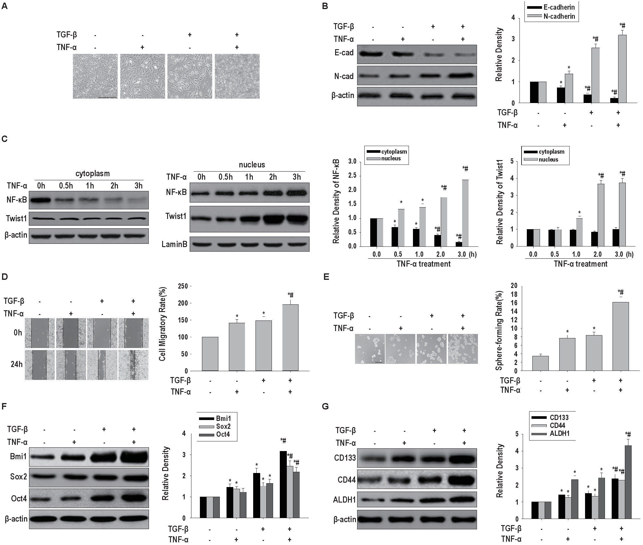
Fig. 1. Co-administration of TGF-β and TNF-α contributes to EMT and CSLC features in HeLa cells. HeLa cells co-administered TNF-α and TGF-β displayed mesenchymal morphology(A, scale bar, 50 μm), reduced E-cadherin and elevated N-cadherin levels(B), increasednuclear translocation of NF-κBp65 and Twist1(C), enhanced migration (D) and self-renewal (E, scale bar, 200 μm) abilities, and upregulated Bmi1, Sox2 and Oct4 (F)as well as CD133, ALDH1 and CD44(H).*p<0.05, vs untreated cells;#p<0.05, vs treated with TNF-α or TGF-β. (C) HeLa cells were treated with TNF-α followed by prolonged induction by TGF-β at different time point. Cytosolic and nuclear NF-κBp65 and Twist1 protein were separated using hypotonic buffer. β-actin and lamin B indicate cytosolic and nuclear fraction, respectively.